Nanoparticle Drug Delivery Systems: Liposomes and Polymeric Nanoparticles

A drug candidate that works perfectly in a test tube can still fail in the body simply because it never reaches its target tissue intact, in sufficient concentration, or without triggering an immune response along the way. Nanoparticle drug delivery systems exist to solve exactly that problem, wrapping a therapeutic payload inside a liposome or polymeric nanoparticle engineered to protect it, extend its circulation time, and in some cases steer it toward diseased tissue while sparing healthy cells. The stakes of getting this right are enormous — nanoparticle platforms now underpin some of the most consequential pharmaceutical products of the last decade, from liposomal chemotherapy to the lipid nanoparticle mRNA vaccines that reached global scale in record time. This guide walks through how liposomes and polymeric nanoparticles differ structurally, how they are manufactured and loaded with drug, how passive and active targeting actually work, and what characterization and regulatory hurdles stand between a bench-scale formulation and a commercial nanomedicine product. For pharmaceutical teams evaluating whether a nanoparticle drug delivery system is the right platform for a molecule, understanding these mechanics is the difference between an informed formulation strategy and an expensive false start.
Liposomes vs Polymeric Nanoparticles: Structure and Composition
Every nanoparticle drug delivery program begins with a platform decision, and that choice determines nearly everything that follows — how the drug is loaded, how it is released, how the immune system responds to the carrier, and how the finished product must be manufactured and stored. Liposomes, polymeric nanoparticles, and lipid nanoparticles are structurally distinct carrier systems built from different materials and self-assembly mechanisms, not variations on a single underlying design. This distinction sits at the technical core of pharmaceuticals and health care innovation, spanning oncology, vaccines, and gene therapy programs alike.
| Platform | Core Material | Typical Payload / Use Case |
|---|---|---|
| Liposome | Phospholipid bilayer vesicle with an aqueous core, often cholesterol-stabilized | Water-soluble and lipophilic small molecules; established chemotherapy platform |
| Polymeric nanoparticle | Solid biodegradable polymer matrix, most commonly PLGA | Sustained-release small molecules and peptides via polymer degradation |
| Lipid nanoparticle (LNP) | Ionizable lipid, helper phospholipid, cholesterol, and PEGylated lipid | Nucleic acids — mRNA and siRNA — via electrostatic complexation |
Biologic drugs face a related but distinct stability challenge before they ever reach a nanoparticle carrier, a subject covered in depth in our guide to biologics formulation and protein stability. Once a platform is selected here, the next design question becomes practical: which manufacturing process can actually produce that structure reliably and at the particle size the therapeutic application demands.
Manufacturing Methods: From Thin-Film Hydration to Microfluidics
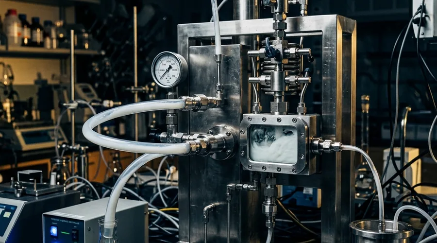
How a nanoparticle is made is inseparable from what it becomes — the manufacturing method directly sets particle size, size distribution, and structural uniformity, and no amount of downstream processing fully corrects a poorly controlled formation step. Each platform has established manufacturing routes, and modern nanomedicine has increasingly shifted toward continuous, precisely controlled processes over older batch methods as particle size control requirements have tightened.
- Thin-film hydration — the classical liposome method, where lipids are dissolved in organic solvent, dried into a film, then hydrated and dispersed into vesicles, typically followed by extrusion or sonication to reduce size
- Solvent evaporation / nanoprecipitation — polymer and drug are dissolved in a water-miscible solvent, then added to an aqueous phase where the polymer precipitates into nanoparticles as the solvent diffuses out
- High-pressure homogenization and extrusion — forces a coarse lipid or polymer dispersion through narrow channels or membranes under pressure to shear particles down to a target diameter
- Microfluidic mixing — rapidly combines an organic lipid stream with an aqueous stream inside a microfluidic chip, the dominant method for producing lipid nanoparticles with tight, reproducible size control
Selecting a manufacturing route settles how a particle is physically built, but it does not yet answer the question that determines clinical performance: how much drug actually ends up inside the particle, and at what rate it comes back out.
Drug Loading, Encapsulation Efficiency, and Release Kinetics
A nanoparticle that forms perfectly but fails to carry a meaningful drug payload is not a viable delivery system, which makes loading strategy and encapsulation efficiency central formulation variables rather than afterthoughts measured only at the end of development. Loading approach differs meaningfully between platforms and depends heavily on the drug's own physicochemical properties — its solubility, ionization state, and log P all determine which loading strategy will actually work.
- Passive loading — the drug is present during particle formation and becomes trapped as the liposome or polymer matrix assembles around it, generally simpler but often lower-efficiency for hydrophilic drugs
- Active (remote) loading — used for liposomes carrying ionizable drugs, exploiting a pH or ion gradient across the lipid bilayer to drive drug accumulation into the aqueous core after the empty liposome has already formed, achieving substantially higher encapsulation efficiency
- Matrix dispersion — for polymeric nanoparticles, the drug is distributed throughout the solid polymer during nanoprecipitation, with release governed by subsequent polymer erosion and diffusion
- Surface adsorption — drug bound to the particle's outer surface rather than encapsulated within it, which tends to produce a faster initial burst release before slower core release begins
Release kinetics from a polymeric nanoparticle trace back to PLGA's well-characterized bulk hydrolysis behavior, in which ester bonds break down into lactic and glycolic acid monomers cleared through the body's normal metabolic pathways, a mechanism reviewed extensively in the pharmaceutical sciences literature on PLGA degradation. Liposomal release, by contrast, depends on bilayer permeability and structural integrity, which is why cholesterol content and lipid phase transition temperature are formulation variables formulators tune deliberately rather than incidentally. Parenteral products built on either platform must also satisfy the sterility and stability demands common to all injectables, covered in our guide to parenteral formulation development.
Getting loading and release chemistry right determines how much drug reaches the body and over what timeframe, but neither variable says anything yet about whether that drug actually arrives where it is needed most — which depends entirely on the particle's targeting strategy.
Targeted Delivery: Passive vs Active Targeting Strategies
Nanoparticles do not automatically know where to go in the body, and directing them toward diseased tissue while avoiding healthy organs is one of the central engineering challenges of the entire field. Two distinct strategies address this, often layered together in a single formulation rather than used in isolation.
- Passive targeting — relies on the enhanced permeability and retention (EPR) effect, in which appropriately sized nanoparticles accumulate preferentially in tumor tissue due to its leaky, abnormal vasculature and poor lymphatic drainage
- PEGylation — coating the particle surface with polyethylene glycol to create a hydrophilic steric barrier that reduces protein opsonization and extends circulation half-life, giving the particle more time to reach its target before clearance
- Active targeting — conjugating ligands such as antibodies, peptides, or small molecules like folate to the particle surface, allowing receptor-mediated binding and uptake specifically by cells overexpressing the corresponding receptor
- Stimuli-responsive release — designing particles that release their payload in response to a local trigger such as tumor-associated pH or specific enzyme activity, adding a further layer of site-selective delivery
Extended circulation time achieved through PEGylation is not without cost: repeated dosing of PEGylated particles has been documented to trigger an anti-PEG antibody response known as the accelerated blood clearance effect, in which subsequent doses clear from circulation faster than the first — a well-established immunogenicity consideration described in the pharmaceutical literature on the EPR effect and its clinical limitations. This is one reason active targeting has gained ground as a complement to, rather than a replacement for, passive EPR-based accumulation.
Once a targeting strategy is designed into the formulation, it has to be proven — not assumed — through rigorous physical characterization before any regulatory or clinical claim can be supported.
Characterization and Quality Control for Nanoparticle Formulations
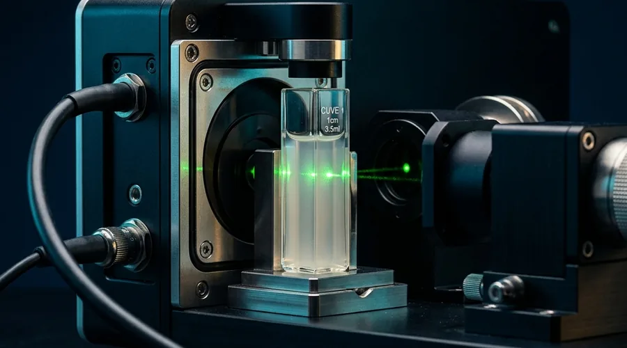
A nanoparticle formulation is only as trustworthy as the data confirming what it actually is — size, charge, and drug content are not cosmetic details but critical quality attributes that determine biological behavior, and each requires a specific analytical method to measure correctly. Nanoparticle characterization is more demanding than most conventional pharmaceutical analysis because the properties that matter most exist at a scale invisible to routine methods.
| Attribute | Method | Why It Matters |
|---|---|---|
| Particle size and PDI | Dynamic light scattering (DLS) | Governs biodistribution, circulation time, and EPR-based accumulation |
| Surface charge | Zeta potential measurement | Determines colloidal stability and interaction with cell membranes |
| Encapsulation efficiency | Free/bound drug separation + HPLC | Confirms how much drug is actually carried per dose, not just formulated |
| Morphology | Transmission electron microscopy (TEM / cryo-TEM) | Directly visualizes structure that light scattering alone cannot confirm |
A tight, well-characterized particle size distribution on paper still has to be reproduced identically batch after batch once a process moves beyond the lab bench, which is where nanoparticle manufacturing runs into its steepest regulatory and scale-up challenges.
Regulatory and Scale-Up Considerations for Nanomedicine
Nanoparticle drug products occupy a regulatory space shaped by their structural complexity — a liposome or lipid nanoparticle is not a simple small-molecule drug substance but a multi-component system whose in vivo performance depends on the physical assembly itself, not just its chemical composition. Regulators evaluate these products accordingly, with chemistry, manufacturing, and controls (CMC) expectations that go well beyond what a conventional oral solid dosage form requires.
In the United States, the FDA has issued specific draft guidance addressing liposome drug products, covering chemistry, manufacturing, controls, and the human pharmacokinetic and bioavailability data expected to support a submission. Nanoparticle manufacturing also carries distinct scale-up risk: mixing speed, mixing geometry, and the rate at which organic and aqueous phases combine all directly set particle size, and these parameters behave nonlinearly when moving from a benchtop process to production-scale equipment, often requiring re-engineered mixing hardware rather than simple proportional scaling. This scale-up challenge compounds with the sterile manufacturing requirement most injectable nanoparticle products carry, since particle formation and aseptic processing must both be validated together rather than sequentially.
Building CMC and scale-up strategy alongside formulation development from the earliest stages, rather than treating it as a downstream manufacturing problem, is what separates nanoparticle programs that reach commercial approval from those that stall after a promising bench-scale result.
Frequently Asked Questions
What's the fundamental difference between a liposome and a polymeric nanoparticle?
A liposome is a spherical vesicle built from a phospholipid bilayer that self-assembles in water, enclosing an aqueous core and mimicking a cell membrane's structure, which makes it well suited to carrying both water-soluble drugs in its core and lipophilic drugs within the bilayer itself. A polymeric nanoparticle, in contrast, is a solid or matrix-type particle built from a biodegradable polymer such as PLGA, in which the drug is dispersed or dissolved throughout the polymer matrix or adsorbed onto its surface.
Liposomes tend to offer gentler drug loading conditions and better biocompatibility for many biologic payloads, while polymeric nanoparticles generally offer more mechanically robust structures and more tunable, sustained release profiles through polymer degradation kinetics. The choice between them is driven by the drug's physicochemical properties, the required release duration, and the manufacturing scale-up pathway a program can realistically support.
How do lipid nanoparticles (LNPs) differ from traditional liposomes?
Traditional liposomes are built primarily from phospholipids and cholesterol arranged in a bilayer with a defined aqueous core, historically developed to encapsulate small-molecule or protein drugs. Lipid nanoparticles, the platform that carries mRNA in the authorized COVID-19 vaccines, use a related but distinct lipid composition — typically an ionizable lipid, a helper phospholipid, cholesterol, and a PEGylated lipid — assembled through rapid microfluidic mixing rather than classical hydration methods.
The ionizable lipid is the key structural difference: it stays largely neutral at physiological pH for storage stability but becomes positively charged at the lower pH used during formulation, allowing it to electrostatically complex with negatively charged nucleic acid payloads like mRNA or siRNA. This makes LNPs the dominant platform for nucleic acid delivery specifically, while liposomes remain more broadly used across small-molecule and protein therapeutics.
What is the EPR effect and why is it central to nanoparticle cancer therapy?
The enhanced permeability and retention (EPR) effect describes how nanoparticles of an appropriate size range tend to accumulate preferentially in tumor tissue compared to healthy tissue, because tumor vasculature is often abnormally leaky with wide endothelial gaps, while tumors also typically have poor lymphatic drainage that traps particles once they arrive. This passive targeting mechanism was the original rationale behind liposomal chemotherapy products, allowing a cytotoxic drug to concentrate more heavily at the tumor site while reducing exposure to healthy organs compared to the free drug alone.
The EPR effect is not uniform across all tumor types or all patients, however, and its clinical reliability has been an active area of research, which is part of why active targeting strategies using surface-conjugated ligands are increasingly combined with passive EPR-based accumulation rather than relied upon alone.
Why is PEGylation used on nanoparticle surfaces, and does it have downsides?
PEGylation refers to coating a nanoparticle's surface with polyethylene glycol chains, which creates a hydrophilic steric barrier that reduces opsonization — the process by which blood proteins bind to a foreign particle and flag it for rapid clearance by the mononuclear phagocyte system. This extends the nanoparticle's circulation half-life substantially, giving it more time to accumulate at its target site via the EPR effect or active targeting before being cleared.
The tradeoff is a phenomenon called the accelerated blood clearance (ABC) effect, in which repeated administration of PEGylated nanoparticles can trigger an anti-PEG antibody response that causes subsequent doses to be cleared much faster than the first, a documented immunogenicity concern that formulators must account for in multi-dose treatment regimens.
How is drug release from a polymeric nanoparticle controlled?
Drug release from a PLGA-type polymeric nanoparticle is governed primarily by polymer degradation kinetics, which proceed through bulk hydrolysis of the polymer's ester bonds into lactic and glycolic acid monomers that are cleared through normal metabolic pathways. Formulators control release duration by adjusting the lactide-to-glycolide ratio, since glycolide-rich polymers degrade faster than lactide-rich ones, along with polymer molecular weight and the particle's overall porosity and size.
Release also depends on whether the drug is encapsulated within the particle's core, dispersed through the matrix, or adsorbed onto the surface, because surface-associated drug tends to show a faster initial burst release before the slower, degradation-controlled release phase takes over.
What analytical methods are used to characterize nanoparticle formulations?
Dynamic light scattering (DLS) is the standard method for measuring particle size and polydispersity index (PDI), reporting how tightly the particle population clusters around its mean diameter, which matters because a broad size distribution can behave inconsistently in vivo. Zeta potential measurement characterizes the surface charge of the particle, which influences both colloidal stability during storage and the particle's interaction with cell membranes and blood components after administration.
Encapsulation efficiency and drug loading are typically quantified by separating free, unencapsulated drug from the nanoparticle fraction and measuring drug content by HPLC, while techniques such as transmission electron microscopy (TEM) or cryo-TEM provide direct visual confirmation of particle morphology that light scattering methods alone cannot supply.
What makes nanoparticle manufacturing difficult to scale from lab to commercial production?
Nanoparticle formation is highly sensitive to mixing speed, mixing geometry, and the rate at which solvent and aqueous phases combine, since these parameters directly determine particle size and size distribution — variables that change nonlinearly when moving from a benchtop process to a larger production vessel or a continuous microfluidic line. A process validated at milliliter scale using manual pipetting or a small microfluidic chip often cannot simply be scaled proportionally, because achieving the same rapid, controlled mixing at liters-per-minute throughput requires re-engineering the mixing hardware itself rather than just running the same process longer.
This is compounded by the need for sterile manufacturing for most injectable nanoparticle products, tight in-process control of particle size and PDI at every batch, and the analytical burden of characterizing complex multi-component particles consistently — all of which is why nanoparticle CMC packages tend to require earlier and more intensive process development investment than conventional small-molecule formulations.
Developing a Nanoparticle Drug Delivery Product?
Global Formulation provides pharmaceutical formulation consultancy, liposomal and polymeric nanoparticle design, CMC and scale-up strategy, and contract development support for nanomedicine programs worldwide.
Talk to Our Formulation Team